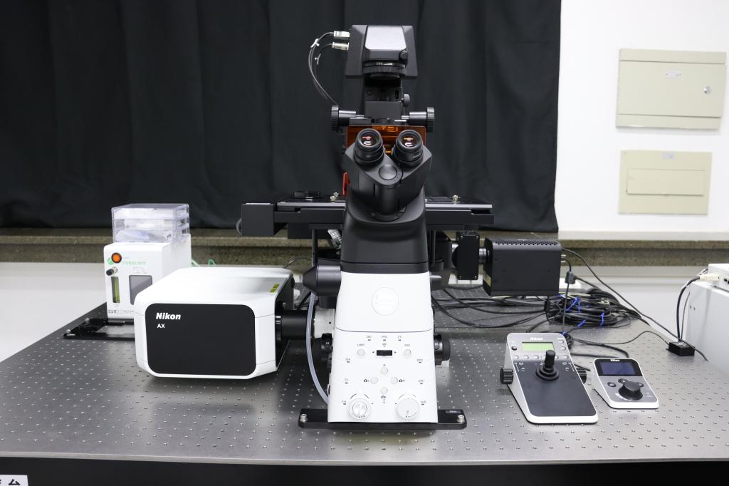
Instrument model:A1HD25
Manufacturer:Nikon
Specifications and technical parameters:1. laser light source: four solid state lasers, laser wavelengths of 405nm, 488nm, 561nm and 640nm, respectively.2. detection channels: with two large target surface PMT and two high-sensitivity GaAsp channels and a transmission imaging channel, the system has a total of five channels, while four-color imaging.3. scanning field of view FOV: field of view diagonal 25mm.4. super-resolution imaging module: X-Y plane imaging resolution of 120nm, Z-axis longitudinal imaging resolution of 300nm.5. flat field compound achromatic high numerical aperture 2x, 10x, 20x, 40x, 60x (oil lens), 100x (oil lens) objective lens.6. Live cell device set For premixed (5% CO2/ 95% air) gas cylinder; with small CO2culture system, control CO2concentration, 37 degrees temperature control, humidity control, 35mm culture dish, coverslip slide culture dish, etc.7. Multi-functional measurement and analysis software, which can analyze and process the image in depth, with automatic measurement, counting and statistical functions, 3D reconstruction function, 3D spatial measurement function.
Features and functions:The equipment is mainly used for the observation and detection of microscopic morphology and microstructure of conventional living cells, biological tissue samples, chemical and nano-material samples. The instrument has the following features:
1. This super-resolution confocal microscope not only has a resolution close to 120nm in XY direction, which elevates the study to the level of organelles and subcellular organelles; but also has a resolution of 300nm in Z-axis, thus not only limited to two-dimensional plane, but also can carry out automated three-dimensional information reconstruction.
2.The machine scanning imaging field of view can reach 25mm field of view, can be a single imaging to obtain more sample information, can greatly improve the efficiency of high-throughput experiments.
3.Introduced a lateral single objective microscope module, mainly for microfluidic motion observation and imaging in the Z-axis direction of the chip.
Attachments and Configurations:1. equipped with 405nm, 488nm, 561nm and 640nm four-wavelength solid-state lasers; 2. equipped with up to 8192X8192 ultra-high resolution scanning head; 3. equipped with fully electric inverted fluorescence microscope. 4. 2-100 times compound achromatic confocal special objective; 5. live cell culture system; 6. professional version of analysis software; 7. high configuration workstation.
Reservation Website:http://202.113.64.51/genee/